3. Bone Structure
Bone tissue (osseous tissue) differs greatly from other tissues in the body. Bone is hard and many of its functions depend on that characteristic hardness. Later discussions in this chapter will show that bone is also dynamic in that its shape adjusts to accommodate stresses. This section will examine the gross anatomy of bone first and then move on to its histology.
Gross Anatomy of Bones
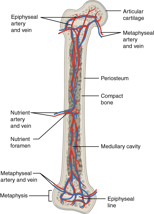
A long bone has two main regions: the diaphysis and the epiphysis (see Figure 3.1). The diaphysis is the hollow, tubular shaft that runs between the proximal and distal ends of the bone. Inside the diaphysis is the medullary cavity, which is filled with yellow bone marrow in an adult. The outer walls of the diaphysis (cortex, cortical bone) are composed of dense and hard compact bone, a form of osseous tissue.
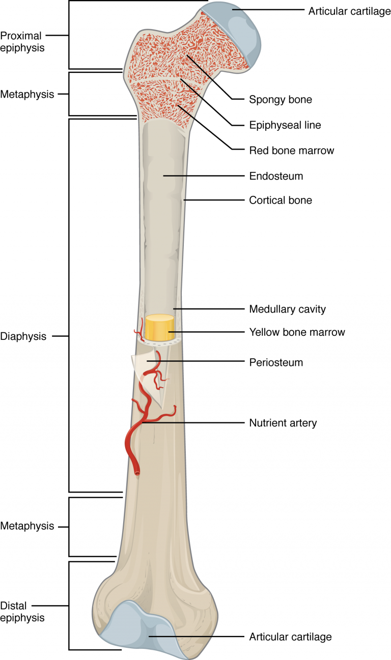
The wider section at each end of the bone is called the epiphysis (plural = epiphyses), which is filled internally with spongy bone, another type of osseous tissue. Red bone marrow fills the spaces between the spongy bone in some long bones. Each epiphysis meets the diaphysis at the metaphysis. During growth, the metaphysis contains the epiphyseal plate, the site of long bone elongation described later in the chapter. When the bone stops growing in early adulthood (approximately 18–21 years), the epiphyseal plate becomes an epiphyseal line seen in the figure.
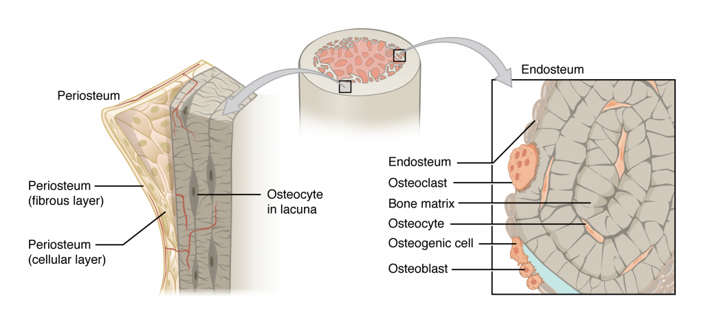
Lining the inside of the bone adjacent to the medullary cavity is a layer of bone cells called the endosteum (endo- = “inside”; osteo- = “bone”). These bone cells (described later) cause the bone to grow, repair, and remodel throughout life. On the outside of bones there is another layer of cells that grow, repair and remodel bone as well. These cells are part of the outer double layered structure called the periosteum (peri– = “around” or “surrounding”). The cellular layer is adjacent to the cortical bone and is covered by an outer fibrous layer of dense irregular connective tissue (see Figure 3.4). The periosteum also contains blood vessels, nerves, and lymphatic vessels that nourish compact bone. Tendons and ligaments attach to bones at the periosteum. The periosteum covers the entire outer surface except where the epiphyses meet other bones to form joints (see Figure 3.2). In this region, the epiphyses are covered with articular cartilage, a thin layer of hyaline cartilage that reduces friction and acts as a shock absorber.
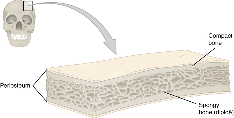
Flat bones, like those of the cranium, consist of a layer of diploë (spongy bone), covered on either side by a layer of compact bone (see Figure 3.3). The two layers of compact bone and the interior spongy bone work together to protect the internal organs. If the outer layer of a cranial bone fractures, the brain is still protected by the intact inner layer.
Osseous Tissue: Bone Matrix and Cells
Bone Matrix
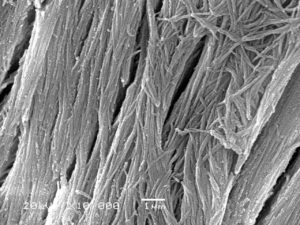
Osseous tissue is a connective tissue and like all connective tissues contains relatively few cells and large amounts of extracellular matrix. By mass, osseous tissue matrix consists of 1/3rd collagen fibers and 2/3rds calcium phosphate salt. The collagen provides a scaffolding surface for inorganic salt crystals to adhere (see Figure 3.4). These salt crystals form when calcium phosphate and calcium carbonate combine to create hydroxyapatite. Hydroxyapatite also incorporates other inorganic salts like magnesium hydroxide, fluoride, and sulfate as it crystallizes, or calcifies, on the collagen fibers. The hydroxyapatite crystals give bones their hardness and strength, while the collagen fibers give them a framework for calcification and gives the bone flexibility so that it can bend without being brittle. For example, if you removed all the organic matrix (collagen) from a bone, it would crumble and shatter readily. Conversely, if you remove all the inorganic matrix (minerals) from bone and leave the collagen, the bone becomes overly flexible and cannot bear weight.
Bone Cells
Although bone cells compose less than 2% of the bone mass, they are crucial to the function of bones. Four types of cells are found within bone tissue: osteoblasts, osteocytes, osteogenic cells, and osteoclasts (see Figure 3.5).
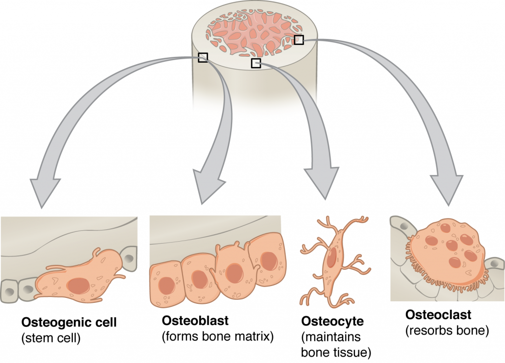
If osteoblasts and osteocytes are incapable of mitosis, then how are they replenished when old ones die? The answer lies in the properties of a third category of bone cells—the osteogenic (osteoprogenitor) cell. These osteogenic cells are undifferentiated with high mitotic activity and they are the only bone cells that divide. Immature osteogenic cells are found in the cellular layer of the periosteum and the endosteum. They differentiate and develop into osteoblasts.
The dynamic nature of bone means that new tissue is constantly formed, and old, injured, or unnecessary bone is dissolved for repair or for calcium release. The cells responsible for bone resorption, or breakdown, are the osteoclasts. These multinucleated cells originate from monocytes and macrophages, two types of white blood cells, not from osteogenic cells. Osteoclasts are continually breaking down old bone while osteoblasts are continually forming new bone. The ongoing balance between osteoblasts and osteoclasts is responsible for the constant but subtle reshaping of bone. Table 2 reviews the bone cells, their functions, and locations.
| Bone Cells (Table 2) | ||
|---|---|---|
| Cell type | Function | Location |
| Osteogenic cells | Develop into osteoblasts | Endosteum, cellular layer of the periosteum |
| Osteoblasts | Bone formation | Endosteum, cellular layer of the periosteum, growing portions of bone |
| Osteocytes | Maintain mineral concentration of matrix | Entrapped in matrix |
| Osteoclasts | Bone resorption | Endosteum, cellular layer of the periosteum, at sites of old, injured, or unneeded bone |
Compact and Spongy Bone
Most bones contain compact and spongy osseous tissue, but their distribution and concentration vary based on the bone’s overall function. Although compact and spongy bone are made of the same matrix materials and cells, they are different in how they are organized. Compact bone is dense so that it can withstand compressive forces, while spongy bone (also called cancellous bone) has open spaces and is supportive, but also lightweight and can be readily remodeled to accommodate changing body needs.
Bone
Compact bone is the denser, stronger of the two types of osseous tissue (see Figure 3.6). It makes up the outer cortex of all bones and is in immediate contact with the periosteum. In long bones, as you move from the outer cortical compact bone to the inner medullary cavity, the bone transitions to spongy bone.
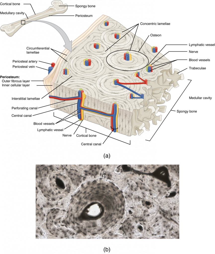
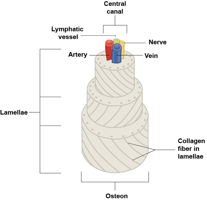
If you look at compact bone under the microscope, you will observe a highly organized arrangement of concentric circles that look like tree trunks. Each group of concentric circles (each “tree”) makes up the microscopic structural unit of compact bone called an osteon (this is also called a Haversian system). Each ring of the osteon is made of collagen and calcified matrix and is called a lamella (plural = lamellae). The collagen fibers of adjacent lamallae run at perpendicular angles to each other, allowing osteons to resist twisting forces in multiple directions (see Figure 3.4). Running down the center of each osteon is the central canal, or Haversian canal, which contains blood vessels, nerves, and lymphatic vessels. These vessels and nerves branch off at right angles through a perforating canal, also known as Volkmann’s canals, to extend to the periosteum and endosteum. The endosteum also lines each central canal, allowing osteons to be removed, remodeled and rebuilt over time.
The osteocytes are trapped within their lacuane, found at the borders of adjacent lamellae. As described earlier, canaliculi connect with the canaliculi of other lacunae and eventually with the central canal. This system allows nutrients to be transported to the osteocytes and wastes to be removed from them despite the impervious calcified matrix.
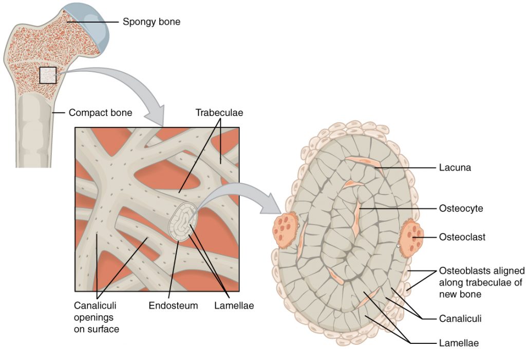
Spongy (Cancellous) Bone
Like compact bone, spongy bone, also known as cancellous bone, contains osteocytes housed in lacunae, but they are not arranged in concentric circles. Instead, the lacunae and osteocytes are found in a lattice-like network of matrix spikes called trabeculae (singular = trabecula) (see Figure 3.8). The trabeculae are covered by the endosteum, which can readily remodel them. The trabeculae may appear to be a random network, but each trabecula forms along lines of stress to direct forces out to the more solid compact bone providing strength to the bone. Spongy bone provides balance to the dense and heavy compact bone by making bones lighter so that muscles can move them more easily. In addition, the spaces in some spongy bones contain red bone marrow, protected by the trabeculae, where hematopoiesis occurs.
Blood and Nerve Supply
The spongy bone and medullary cavity receive nourishment from arteries that pass through the compact bone. The arteries enter through the nutrient foramen (plural = foramina), small openings in the diaphysis (see Figure 3.9). The osteocytes in spongy bone are nourished by blood vessels of the periosteum that penetrate spongy bone and blood that circulates in the marrow cavities. As the blood passes through the marrow cavities, it is collected by veins, which then pass out of the bone through the foramina.
In addition to the blood vessels, nerves follow the same paths into the bone where they tend to concentrate in the more metabolically active regions of the bone. The nerves sense pain, and it appears the nerves also play roles in regulating blood supplies and in bone growth, hence their concentrations in metabolically active sites of the bone.
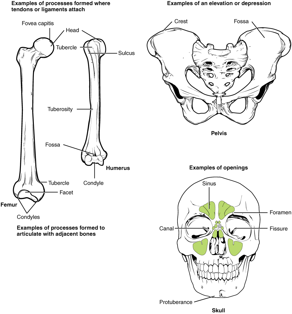
Bone Markings
Define and list examples of bone markings
The surface features of bones vary considerably, depending on the function and location in the body. Table 3 describes the bone markings, which are illustrated in (see Figure 3.10). There are three general classes of bone markings: (1) articulations, (2) projections, and (3) holes. As the name implies, an articulation is where two bone surfaces come together (articulus = “joint”). These surfaces tend to conform to one another, such as one being rounded and the other cupped, to facilitate the function of the articulation. A projection is an area of a bone that projects above the surface of the bone. These are the attachment points for tendons and ligaments. In general, their size and shape is an indication of the forces exerted through the attachment to the bone. A hole is an opening or groove in the bone that allows blood vessels and nerves to enter the bone. As with the other markings, their size and shape reflect the size of the vessels and nerves that penetrate the bone at these points.
| Bone Markings (Table 3) | ||
|---|---|---|
| Marking | Description | Example |
| Articulations | Where two bones meet | Knee joint |
| Head | Prominent rounded surface | Head of femur |
| Facet | Flat surface | Vertebrae |
| Condyle | Rounded surface | Occipital condyles |
| Projections | Raised markings | Spinous process of the vertebrae |
| Protuberance | Protruding | Chin |
| Process | Prominence feature | Transverse process of vertebra |
| Spine | Sharp process | Ischial spine |
| Tubercle | Small, rounded process | Tubercle of humerus |
| Tuberosity | Rough surface | Deltoid tuberosity |
| Line | Slight, elongated ridge | Temporal lines of the parietal bones |
| Crest | Ridge | Iliac crest |
| Holes | Holes and depressions | Foramen (holes through which blood vessels can pass through) |
| Fossa | Elongated basin | Mandibular fossa |
| Fovea | Small pit | Fovea capitis on the head of the femur |
| Sulcus | Groove | Sigmoid sulcus of the temporal bones |
| Canal | Passage in bone | Auditory canal |
| Fissure | Slit through bone | Auricular fissure |
| Foramen | Hole through bone | Foramen magnum in the occipital bone |
| Meatus | Opening into canal | External auditory meatus |
| Sinus | Air-filled space in bone | Nasal sinus |